Home

Solutions

Oncology Med Bot CareLinq AI Pharma Marketing AI NuGleason PathVu Computer Vision AI

Company

About Security Insights Contact us Request a demo
Digital pathology · IVF · Image integrity

Computer Vision AI: digital pathology AI platform for laboratories and clinics

See beyond, decide better. AI-driven image analysis for clinical excellence.

Primary target: digital pathology AI platform

What it is

Three image workloads on one platform

Computer Vision AI is the digital pathology AI platform from PRR.AI. It runs three image workloads: pathology AI for cancer detection and grading on whole slide images, IVF intelligence for embryo assessment, and fraud detection for medical image and report integrity. Two named products sit on it: NuGleason for Gleason grading of prostate cancer, and PathVu for whole slide image viewing and collaboration.

Every clinical output uses a human-in-the-loop design: the AI proposes, the pathologist or embryologist corrects in real time, and the correction is recorded.

01

Optimises clinical decision-making

Models return real-time insight to the clinician to support accurate grading and embryo selection.

02

Streamlines the laboratory workflow

Integration with pathology labs and clinics automates handling steps and reduces the manual load on the reporting queue.

03

Improves throughput without losing the check

Time saved on routine reads goes back into the cases that need a second opinion.

Modules

What the platform does, module by module

Each module below serves a different buyer within your organization.

Pathology AI

For the pathology laboratory head and the reporting pathologist.

Digital slide processing

AI-driven annotation, measurement and automated diagnostics on pathology images.

Histopathology workflow management

A centralised platform with real-time insight and collaboration tools across the reporting team.

AI-powered cancer grading

Models for prostate, breast and other cancer grading, delivered with heatmaps and structured scores.

Human-in-the-loop

AI-assisted decision support with real-time error correction by the reporting pathologist.

IVF intelligence

For the IVF clinic director and the embryology team.

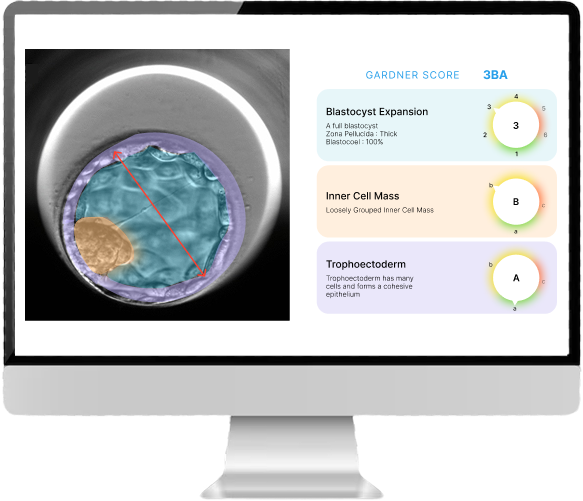
AI-powered Gardner scoring

Evaluates embryo quality by analysing expansion, inner cell mass and trophectoderm, against the Gardner grading system embryologists already use.

Real-time clinical insight

Supports the embryologist's own assessment rather than replacing it, so selection decisions stay with the clinic.

AI-powered fraud detection

For the payer, the TPA and the hospital audit function.

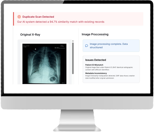
Duplicate scan detection

Identifies reused radiology images such as ultrasound, CT and MRI using deep image comparison and perceptual hashing.

Metadata mismatch detection

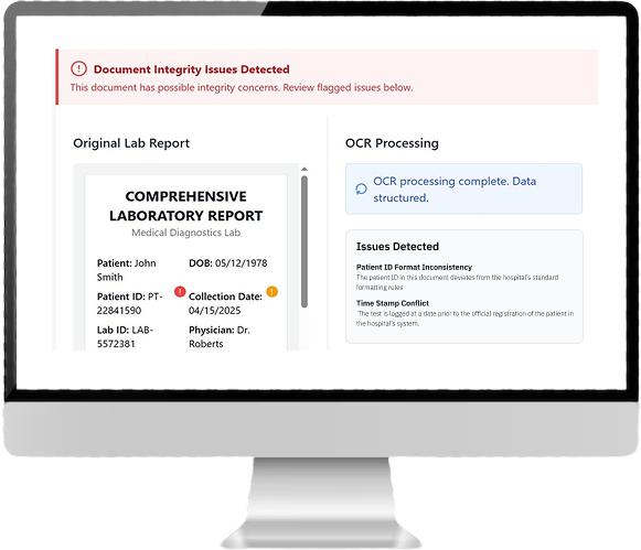
Extracts and analyses timestamps, patient IDs and scan metadata with OCR and NLP to flag inconsistencies.

Smart document forensics

Flags fabricated reports from font, layout and signature evidence, and catches duplicates filed under different IDs.

Data integrity and anomaly scanner

Cross-checks dates, patient IDs and timestamps against claims databases, while NLP detects suspicious narratives and inflated treatments.

Med AI Services

Delivered work rather than licensed software, for teams without an in-house pathology AI function.

Remote tele-pathology

Reporting powered by AI and PRR.AI pathology experts, for laboratories without local subspecialty cover.

Research and trial support

AI-driven tumour segmentation, biomarker analysis and cancer grading, plus model training for research and clinical trials with API integration.

The modules in use

One vision stack, four very different jobs

The same underlying platform reads a prostate biopsy, grades a blastocyst and flags a tampered radiology record. What changes is the model and the report, not the infrastructure.

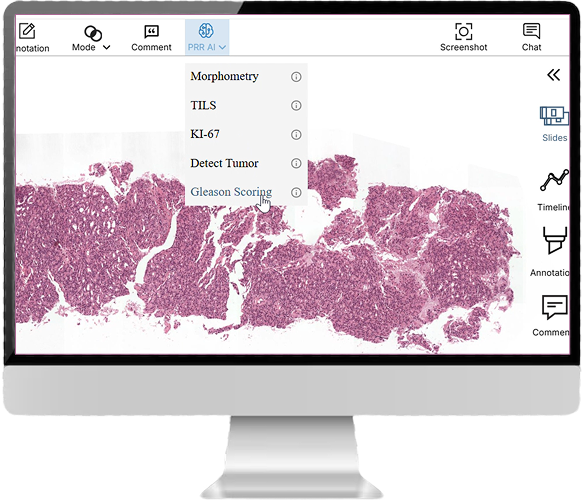
The PRR AI analysis menu open over a whole slide image, listing Morphometry, TILS, KI-67, Detect Tumor and Gleason Scoring.
Digital pathology. Morphometry, TILs, Ki-67, tumour detection and Gleason scoring run from inside the viewer, against the slide already open.
Blastocyst image with the inner cell mass and blastocoel segmented in colour, alongside a Gardner score of 3BA broken into blastocyst expansion 3, inner cell mass B and trophectoderm A.
IVF embryo grading. Gardner scoring split into its three components — expansion, inner cell mass and trophectoderm — each with the segmentation it was derived from.
A duplicate scan warning reporting a 94.7 percent similarity match with existing records, beside the original chest X-ray and a list of detected issues including a patient ID mismatch and EXIF timestamp inconsistency.
Duplicate scan detection. Identical radiographic content filed under two patient IDs, caught by image similarity rather than by metadata alone.
A document integrity warning over a scanned laboratory report, with OCR processing complete and issues detected including a patient ID format inconsistency and a time stamp conflict.
Document integrity. OCR structures the report, then checks the result against the hospital’s own ID formats and registration timestamps.
Named products

NuGleason and PathVu

Each has its own page and its own keyword, so they do not compete with this one in search.

Research

CoNIC Challenge: nuclear segmentation and classification in colon histology

PRR.AI's pathology team entered the Colon Nuclei Identification and Counting (CoNIC) grand challenge, which asked entrants to segment, classify and count six nucleus types across the largest publicly available nuclei-level dataset in computational pathology.

  • Six nucleus classes: epithelial, lymphocyte, plasma, eosinophil, neutrophil, connective tissue
  • Lizard dataset, H&E-stained histology at 20x, around half a million labelled nuclei
  • 238 images split 204 training and 34 validation, patched at 244x244
  • Hematoxylin-aware dual-encoder architecture with PDFA blocks over a Preact-ResNet backbone

Method write-up published on prr.ai, December 2022.

Questions

Computer Vision AI: frequently asked questions

What is Computer Vision AI?

Computer Vision AI is the digital pathology AI platform from PRR.AI. It runs pathology AI for cancer detection and grading on whole slide images, IVF intelligence for embryo assessment, and fraud detection for medical image and report integrity.

Which cancers can the platform grade?

The platform includes models for prostate, breast and other cancer grading. Prostate grading is delivered as the named product NuGleason, which returns heatmaps, Gleason scores and standardised reports.

How does the human-in-the-loop approach work?

The AI proposes a segmentation, a grade or a score, and the reporting pathologist corrects it in real time inside the workflow. The correction is recorded. The pathologist signs the report, not the model.

Does the platform do IVF embryo grading?

Yes. IVF intelligence applies AI-powered Gardner scoring, evaluating embryo expansion, inner cell mass and trophectoderm to support the embryologist's assessment. Embryo selection remains a clinic decision.

Is Computer Vision AI a diagnostic device?

No. The pathology and IVF modules are decision support used with a clinician in the loop, and they are not stand-alone diagnostic devices. Regulatory status differs by module and by market, so ask us for the current position for yours.

What scanners and file formats does it support?

The platform reads whole slide images and integrates with laboratory information systems and image stores.
Computer Vision AI

Bring us ten slides

Send a small, de-identified set from your own scanner. We will run it and walk you through the output with your pathologist.

Get started